Mineralized scale patterns on the cell periphery of the chrysophyte Mallomonas determined by comparative 3D Cryo-FIB SEM data processing
M. Hörning, A. Schertel, R. Schneider, M.-L. Lemloh, M. R. Schweikert, and I. M. Weiss
Journal for Structural Biology, 209(1), 107403, 2020
doi : 10.1016/j.jsb.2019.10.005
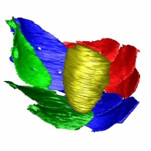
Brief Summary: ''... here the scale case of the photosynthetic synurophyte Mallomonas was preserved in aqueous suspension using high-pressure freezing (HPF). From this specimen, a three-dimensional (3D) data set was collected by Cryo-FIB SEM. SEM imaging using In-lens SE detection allowed to clearly differentiate between mineralized, curved scales of less than 0.2μm thickness and organic cellular ultrastructure or vitrified ice. The three-dimensional spatial orientations and shapes of a minimum set of scales (N=13) were identified, and two segmentation approaches were comparatively applied. Computational automated routines and principal component analysis of the experimentally extracted data created a realistic mathematical model based on the Fibonacci pattern theory. ''