High-resolution imaging techniques have shown that many ion channels are not static, but are subject to highly dynamic processes that include transient association of pore-forming and auxiliary subunits, lateral diffusion and clustering with other proteins, which in turn affects their function 1-3. However, the link between lateral diffusion and function is poorly understood.
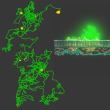
In the paper, recently published by Shuo Wang et al. (for reference, see here), we used a novel electrode-free optical model system 4 to simultaneously monitor and subsequently correlate lateral mobility and single channel activity of a multisubunit membrane protein complex (TOM-CC, also known as the general protein import pore of the mitochondria 5-9) in membranes resting on ultrathin hydrogel films. This optical approach relies on Ca 2+-ion flux through individual channels, which is measured by observing the fluorescence emission of a Ca 2+-sensitive dye in close proximity to the membrane using total interference reflection fluorescence (TIRF) microscopy. In contrast to classical approaches of single-molecule tracking, no fluorescent fusion proteins or labels, which might interfere with lateral movement and function in the membrane, are required. Possible changes in ion flux associated with protein conformational changes are only caused by thermal motion of the protein and possible interactions with the hydrogel underlying the membrane.
We found that freely diffusing TOM-CC molecules stall when interacting with structures adjacent to the membrane, ostensibly due to interaction between its extended polar domains and the supporting hydrogel film. Surprisingly, however, and concomitantly with suspension of movement, TOM-CC changes reversibly from an active (fully light, S H) to a weakly active (medium light, S I) and inactive (dark, S L) channel state. The strong temporal correlation between lateral mobility and ion permeability suggests that TOM-CC channel gating is highly sensitive to molecular confinement and the mode of lateral diffusion. What causes the transitions between the open and the closed conformations of the two TOM-CC pores? What is acting as the “light-intensity-switch”?
Taken alongside recent cryoelectron microscopy of the TOM-CC 5-9, we argue that the two Tom22 subunits of TOM-CC can force the two pore-forming β-barrel Tom40 subunits to undergo a conformational change that leads to channel closure, and that this dynamic effect represents a new functionality of TOM-CC. It will be interesting to study as to whether the mechanostimulated conformational change of the TOM-CC observed in vitro can be confirmed in intact mitochondria, where mechanosensitive interactions with components of the mitochondrial inner membrane are of physiological importance. In this context, it might also be interesting to visualize different Tom22-dependent conformations of TOM-CC using cryo-EM, and to perform single-molecule FRET experiments to study the dynamics of TOM-CC conformers. From a general perspective, to the best of our knowledge, we believe that this may be the first demonstration of mechanoregulation of a β-barrel membrane protein complex and the causal effect of lateral diffusion. So far, mechanosensitivity has only be reported for α-helical proteins 10-12.
We believe that the experimental approach we have used can be readily applied to other mechanosensitive channels where the lateral mobility, e.g. free, restricted or directed diffusion, of integral and peripheral membrane proteins is an important factor in the organisation and function of biological membranes and their components. Our study also underlies the importance of the bottom-up approach, using reconstituted systems, to understand interactions in complex physiological environments.
"Behind the paper" blog post:
Wang, S. & Nussberger, S. Optical single-channel recordings reveal correlation between lateral protein diffusion and channel activity of TOM. Shuo Wang et al. Nature Portfolio Bioengineering Community (2022)
References:
- Kusumi, A., Tsunoyama, T. A., Hirosawa, K. M., Kasai, R. S. & Fujiwara, T. K. Tracking single molecules at work in living cells. Nat. Chem. Biol. 10, 524–532 (2014).
- Heine, M., Ciuraszkiewicz, A., Voigt, A., Heck, J. & Bikbaev, A. Surface dynamics of voltage gated ion channels. Channels 10, 267–281 (2016).
- Jacobson, K., Liu, P. & Lagerholm, B. C. The lateral organization and mobility of plasma membrane components. Cell 177, 806–819 (2019).
- Leptihn, S. et al. Constructing droplet interface bilayers from the contact of aqueous droplets in oil. Nat. Protoc . 8, 1048–1057 (2013).
- Ahting, U., Thun, C., Hegerl, R., Typke, D., Nargang, F.E., Neupert, W., & Nussberger, S. The TOM core complex: the general protein import pore of the outer membrane of mitochondria. J. Cell Biol. 147, 959–968 (1999).
- Bausewein, T., Mills, D.J., Langer, J.D., Nitschke, B., Nussberger, S. & Kühlbrandt, W. Cryo-EM structure of the TOM core complex from Neurospora crassa. Cell 170, 693–700 (2017).
- Araiso, Y. et al. Structure of the mitochondrial import gate reveals distinct preprotein paths. Nature 575, 395–401 (2019).
- Tucker, K. & Park, E. Cryo-EM structure of the mitochondrial protein-import channel TOM complex at near-atomic resolution. Nat. Struct. Mol. Biol. 26, 1158–1166 (2019).
- Wang, W. et al. Atomic structure of human TOM core complex. Cell Discov. 6, 1–1 0 (2020).
- Martinac, B. Mechanosensitive ion channels: molecules of mechanotransduction. J. Cell Sci. 117, 2449–2460 (2004)
- Jin, P., Jan, L. Y. & Jan, Y.-N. Mechanosensitive ion channels: structural features relevant to mechanotransduction mechanisms. Annu. Rev. Neurosci. 43, 207–229 (2020).
- Kefauver, J. M., Ward, A. B. & Patapoutian, A. Discoveries in structure and physiology of mechanically activated ion channels. Nature 587, 567–576 (2020).